A new examination of the prostate with suspicion of prostate carcinoma is performed. Therefore, an MRI of the prostate is generated and sent to the radiology PACS.

PAIRADS is a research project in the field of the integration of artificial intelligence in radiology. The main goal is to develop the best possible integrability and a demonstrator for the detection of prostate carcinomas. This has already resulted in the following core features of the project.
The human-in-the-loop approach describes the inclusion of, for example, radiologists in the learning process of the system. The radiologists can be seen as mentors for the artificial intelligence that improve the results of the system.
The technology of Artificial Intelligence as well as its results is intangible for many. Therefore, we focus on an approach to make the results of AI better understandable, which is called Explainable AI. In doing so, we relied on both graphical and textual explanations.
We rely on proven methods for the detection of prostate carcinomas, such as the PI-RADS standard. In doing so, we are working with domain experts to develop a system that works according to known characteristics of the PI-RADS standard.
A new examination of the prostate with suspicion of prostate carcinoma is performed. Therefore, an MRI of the prostate is generated and sent to the radiology PACS.

In order to keep your patients' data protected we already pseudonymize the patient's image data fully automatically in your network. Therefore, only information that is really important for us is sent to PAIRADS. The DICOM-Gateway from Gemedico is used for this.

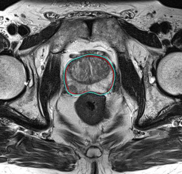
Our system first starts by detecting the prostate within the images. This helps us in further calculations such as mainly for further segmentation of the prostate and calculation of the volume.

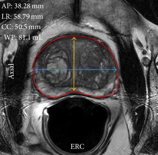
An important feature according to the PI-RADS standard is the volume of the prostate. The previous step allows us to calculate the volume, which is additionally included in the result.

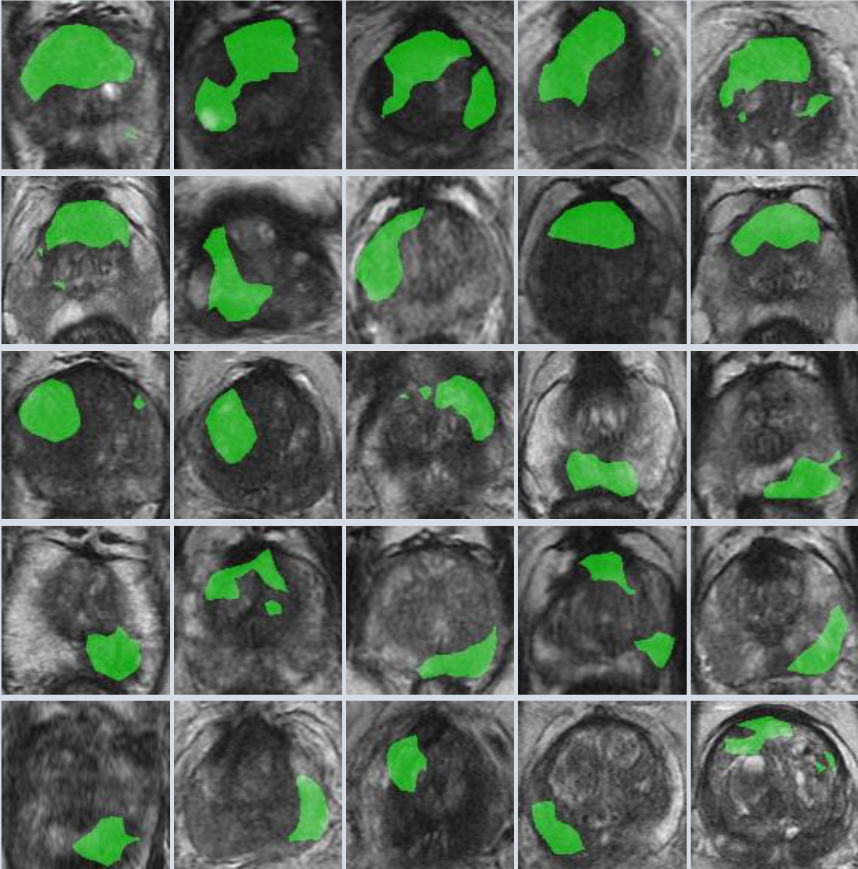
This step is one of the most challenging. The system applies what it has learned to the study and detects prostate carcinomas. However, this does not mean that the system already knows where the carcinoma is located but only that an abnormality exists.

Using Explainable AI, we localize in which section of the prostate an abnormality was found. Combining the segmentation of the prostate, we can tell the radiologist more specifically where we detected an abnormality.

The results of the system are summarized and made available to radiologists. Therefore, segmentations, volumes, carcinomas and its localization are assembled into one package and sent.

The radiologists now have the opportunity to evaluate the results of the system. For example, segmentations of the prostate can be created, carcinomas can be added or removed, or the volume can be adjusted. This step is optional, i.e. the productivity of the radiologists is not restricted by this.

Are you a radiologist interested in new opportunities and would like to help with this project? Please contact us! We are permanently looking for partner radiologists who would like to use our system and help us to adapt the system to the needs of radiologists. The more radiologists support us, the more views we get and we believe that more views make a better system!
You are not a radiologist but you have prostate images that you would like to provide us? Then feel free to contact us! We are very grateful for any study that is made available to us, as it is very difficult to obtain quallitatively good images.
I would like to help

Loading ...